Testing for fatigue
This article is available to download as a PDF version
How pathology testing can help identify the cause of patient fatigue
Fatigue is a symptom which can have debilitating effects on patients, posing a negative impact on their overall quality of life including work, family life and general social relationships. Patients experiencing chronic, ongoing fatigue often find it difficult to convey their symptoms to their healthcare professionals. What if there was a way to test for conditions associated with fatigue, giving practitioners and their patients a clearer more targeted treatment approach? Studies have confirmed that without treatment the prognosis of patients with idiopathic fatigue is poor.

Pathology testing is the first step in identifying the cause of patients’ fatigue. The most common conditions associated with fatigue are usually anaemia, thyroid disorders and vitamin D deficiency. These disorders have a known pathophysiological association with fatigue, therefore testing for these first will provide practitioners with a baseline for potential underlying causes of fatigue.
Stress and poor sleep can also have direct influence in elevated levels of fatigue. Testing patients’ salivary cortisol and melatonin levels can provide further insight into potential contributors to fatigue symptoms.
Gut health and genetics can also be implicated in the development of fatigue symptoms. Testing for intestinal permeability (leaky gut) or identifying a polymorphisms on the MTHFR gene methylation pathway, may be useful particularly with patients presenting with idiopathic symptoms of fatigue.
Detailed below are pathology tests which may prove to be beneficial in establishing underlying causes of fatigue and may assist in monitoring treatment progress.
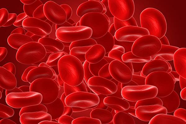
Anaemia
One of the most common conditions associated with fatigue, is anaemia. Anaemia can be caused by a reduced number of red blood cells or impaired haemoglobin production, which is responsible for transporting oxygen in peripheral blood.
As the levels of haemoglobin drop, the ability of the blood to deliver oxygen to organs, muscles and tissues declines. This means that less energy is available to these organs resulting in symptoms perceived as fatigue. This is particularly evident when there is a reduction in cerebral oxygenation. Some studies have suggested that fatigue may in fact be due to deficiency of iron at a cellular level rather than anaemia, as iron plays a major role in the formation of red blood cells.
The varied causes of anaemia require different treatment, therefore it is essential to identify the type of anaemia before any treatment can be initiated.
Available Tests
Identification of anaemia is based on a Full Blood Examination (FBE) and reticulocyte count. This test will clarify the type of anaemia the patient may be presenting with. Too much iron, in conditions such as thalassemia can also be problematic causing similar symptoms of fatigue.
The full iron studies test will also help in identifying whether there may be iron deficiency or excess. Pathology testing is essential to identify the underlying cause of symptoms as the treatment for these conditions would be very different.
Thyroid Insufficiency
Fatigue is a common symptom of thyroid disease. The adrenal and the thyroid systems are closely involved with direct cross regulation between the hypothalamic-pituitary-adrenal (HPA) axis and the hypothalamic-pituitary-thyroid (HPT) axis. Increased stress can cause a reduction in the production of thyroid hormones. Glucocorticoids can inhibit conversion of T4 to T3 resulting in a reduction of serum T3 levels. Studies have also confirmed that prolonged stress can cause a decrease in peripheral thyroid hormone levels.
Thyroid hormone is required in several energy producing processes, namely, the Creb’s citric acid cycle and in oxidative phosphorylation. A thyroid problem can in fact have a direct impact on ATP producing mechanisms thereby resulting in fatigue.

Thyroid hormone Testing
Monitoring thyroid levels in patients presenting with fatigue can provide insight into symptoms that may be stemming from thyroid insufficiency.
Vitamin D deficiency
Low levels of vitamin D have been seen in patients presenting with fatigue. More so, it has been found that normalization of low vitamin D levels in patients with fatigue significantly improves symptoms.
There are a number of mechanisms by which vitamin D is believed to ameliorate symptoms of fatigue. Vitamin D is involved in the process of energy production in the body. Research has shown that low levels of vitamin D are associated with suboptimal mitochondrial function and therefore reduced energy levels. Mitochondria are part of the cell involved in ATP production, the body’s main energy production mechanism.
Vitamin D is also required for calcium homeostasis, supporting bone density and turnover. A deficiency in Vitamin D results in skeletal demineralisation and muscle weakness. Fatigue is believed to be the end result of the underlying muscle fatigue, which is more commonly encountered than muscle weakness.
Additionally, Vitamin D is also involved in supporting immune function by down regulating cytokine, T helper cell and NF-κB activation, therefore reducing pro-inflammatory immune responses, and enhancing immune function.
Testing for Vitamin D
Testing serum vitamin D is a reliable and effective way of monitoring potential vitamin D deficiency. Supplementation can readily rectify deficient vitamin D levels
Adrenal Hormone
Adrenal hormones, predominantly cortisol has been found to have a direct relationship with fatigue. Studies show reduced function of the hypothalamic-pituitary-adrenal (HPA) axis as well as hypocortisolism (reduced cortisol levels, seen via reduced salivary cortisol levels), in people suffering with fatigue.
Available Tests
Testing cortisol levels in saliva is an effective way of monitoring adrenal levels. Saliva is the most reliable way to measure free bioavailable hormone levels at a cellular level, as it reflects the non-protein bound ‘free’ fraction of hormones at a given point in time. Our tests are able to be performed in the privacy and comfort of the patients’ home, therefore allowing for testing at multiple time points throughout the day. Studies have confirmed that fatigue has a diurnal pattern similar to that of cortisol, therefore this method of testing provides an effective way of monitoring cortisol levels throughout the day.
Melatonin dysregulation
A common complaint of patients with fatigue is sleep impairment. The hormone melatonin is significant in the regulation of the circadian rhythm, regulating the sleep-wake cycle.
Ideally in humans melatonin levels should start to increase with the onset of darkness, and reduce again during the second half of the night. This pattern may be altered in patients suffering with fatigue. Studies have found that Melatonin levels are in fact elevated in some people suffering from fatigue. It has been hypothesised that this elevation in melatonin levels may be due to the dysregulation of the hypothalamus-pituitary-adrenal (HPA) axis, however this is yet to be confirmed.

Saliva Melatonin Testing
A simple saliva melatonin test performed at night prior to sleep and again on waking can demonstrate the fluctuations of an individual’s melatonin hormone levels
Gastrointestinal Tract- Leaky Gut
Ongoing fatigue, in particular chronic fatigue syndrome (CFS), can be associated with an immune disorder due to the induction of pro-inflammatory cytokine levels and increased oxidative stress seen in patients with this condition.
Patients with chronic fatigue syndrome are more likely to present with increased levels of IgA where the levels of serum IgA, identified directly correlated with the severity of symptoms. Levels of enterobacteria are also significantly higher in patients with CFS. It is believed that enterobacteria is involved in the aetiology of CFS.

Increased intestinal permeability or ‘leaky gut’ allows the enterobacteria to cross the gut epithelium, increasing pro-inflammatory cytokine production and oxidative stress, resulting in the induction of central neuro-inflammation. Leaky gut can be the underlying cause of CFS allowing enterobacteria species to infiltrate the gut, or may develop as a result of CFS where the activation of pro-inflammatory pathways such as increased cytokine and NFκΒ production may facilitate loosening of tight junctions in the epithelial lining.
It has been shown that fatigue and other symptoms of CFS are reduced with gut permeability treatment.
Available Tests
Patients presenting with fatigue would benefit from monitoring levels of IgA as well as evaluating potential leaky gut through urinary Intestinal Permeability testing, a simple challenge test
MTHFR
Genetic factors and compromised methylation pathways can play a significant role in the predisposition and development of fatigue. Methylation is a process in the body where a methyl group is donated to a molecule, to ensure proper functioning of metabolic pathways. One important methylation process is that of homocysteine. Homoceysteine is remethylated to methionine utilising the enzyme MTHFR (methyltetrahydrofolate reductase). The MTHFR enzymatic pathway utilizes folate to convert to its active form, folinic acid, which is required to facilitate the remethylation of homocysteine to methionine.
A polymorphism in this gene can reduce the efficacy of the MTHFR enzyme, causing reduced conversion of homocysteine to methionine, resulting in elevated levels of homocysteine and reduced activation of folate to its active form.
Vitamin B12 and folate are essential for brain development and function, a deficiency in these nutrients has been linked to an increase in neurological symptoms such as fatigue. Furthermore, this methylation pathway is involved in activation and production of SAMe, an important methyl donor, especially in the brain, which is required for neurotransmitter synthesis. Elevated levels of homocysteine also have a direct adverse effect on neurons and blood vessels including those that are in the brain, contributing to symptoms.
Another common finding associated with a defect of MTHFR is an increase in copper levels. Elevated copper levels have been linked to conditions such as hyperactivity, headaches, depression, increased adrenal stress, fatigue and can also be responsible for a lowered immune system. Elevated copper levels may also make it difficult to raise iron levels, another contributing factor to symptoms of fatigue.
Testing for MTHFR
A simple swab or blood test can determine whether an individual has a mutation on either the C677T and/ or the A498C variants. This test can provide vital information into potential causes of fatigue which may be readily rectified should the MTHFR polymorphism be involved.
Testing for serum B12, folate and homocystein levels show whether the MTHFR polymorphism is affecting the patient. Red Cell copper levels can also be useful in monitoring copper toxicity levels.
There are many reasons patients present to their healthcare professional with fatigue. Pathology testing provides key insights into potential causes so that appropriate and effective treatment can be considered.
BeFunctional prides itself on providing our referrers with the highest levels of scientific expertise and integrity. All of our tests are performed by a highly skilled team of scientific and technical staff. We are committed to providing superior technical support to our practitioners to discuss appropriate test selection, interpretation of test results, review and discussion individual cases and any other related clinical issues.
Contact us today to get your free starter pack, and start referring your patients for pathology testing to get a clearer insight into the underlying causes of their presenting conditions.
References:
Rosenthal, TC, Majeroni, BA, Pretorius, MK 2008, ‘Fatigue: An Overview ‘, American Family Physician, Vol. 78, no. 10, pp. 1173-1180.
Sharpe, M & Wilks, D 2002, ‘Fatigue’, BMJ, Vol. 325, no. 7362, pp. 325-480.
Adrenal Hormone
Jerjes, WK, Clearw, AJ, Wessely, S, Wood, PJ, Taylor, NF 2005, ‘Diurnal patterns of salivary cortisol and cortisone output in chronic fatigue syndrome’, Journal of affective disorders, vol. 87, no. 2-3, pp. 299-304.
Minetto, MA, Lanfranco, F, Tibaudi, A, Ghigo, E 2008, ‘ Changes in awakening cortisol response and midnight salivary cortisol are sensitive markes of strenuous training-induced fatigue’, Journal of endocrinological Investigation, vol. 31, no. 1, pp.16-24.
Roberts, ADL, Papadopoulos, AS, Wessely, S, Chalder, T, Cleare AJ 2009, ‘Salivary cortisol output before and after cognitive behavioural therapy for chronic fatigue syndrome’, Journal of Affective Disorders, vol. 115, no. 1-2, pp.280-286.
Roberts, AD L, Wessely, S, Chalder, T, Papadopoulos 2004, ‘ Salivary cortisol response to awakening in chronic fatigue syndrome’, The British Journal of Psychiatry, vol. 184, no. 2, pp.136-141.
Wolbeek, M, Lorenz, J.P, van Doornen, Coffeng, LE, Kavelaars, A, Heijnen, CJ 2007, ‘ Cortisol and sever fatigue: A longitudinal study in adolescent girls’, Psychoneuroendocrinology, vol. 32, no. 2, pp.171-182.
Melatonin
Knook, L, Kavelaars, A, Sinnema, G, Kuis, W, Heijnen, CJ 2000, ‘ High nocturnal Melatonin in adolescents with chronic fatigue syndrome, The Journal of Clinical Endocrinology & Metabolism, vol. 85, no. 10, pp. 3690-3692.
Lieberman, HR, Waldhauser, F, Garfield, G, Lynch, HJ, Wurtman, RJ 1984, ‘Effects of melatonin on human moon and performance’, Brain Research, vol 323, no. 2, pp. 201-207.
Yancey, JR, Thomas, ST 2012, ‘Chronic Fatigue Syndrome: Diagnosis and Treatment’, American Family Physician, vol. 86, no. 8, pp. 741- 746.
Leaky gut
Maes, M & Coucke, F 2007, ‘Normalization of the increased translocation of endotoxin from gram negative enterobacteria (leaky gut) is accompanied by a remission of chronic fatigue syndrome’, Neuroendocrinology Letters, vol. 28, no. 6, pp.739-744.
Maes, M, Mihaylova, I, Leunis, JC 2007, ‘Increased serum IgA and IgM against LPS of enterobacteria in chronic fatigue syndrome (CFS): Indication for the involvement of gram-negative enterobacteria in the etiology of CFS and for the presence of an increased gut–intestinal permeability’, Journal of Affective Disorders, vol. 99, no. 1-3, pp. 237-240.
Maes, M & Leunis, JC 2008, ‘ Normalisation of leaky gut in chronic fatigue syndrome (CFS) is accompanied by a clinical improvement: effects of age, duration of illness and the translocation of LPS from gram- negative bacteria’, Neuroendocrinology Letters, vol. 29, no. 6, pp. 902-910.
Maes, M 2009, ‘Leaky gut in chronic fatigue syndrome: A review’, Activities Nervosa Superior Rediviva, vol. 51, no. 1-2, pp. 21-28.
Klimas, NG & Koneru, AO 2007, ‘ Chronic fatigue syndrome: Inflammation, immune function, and neuroendocrine interactions’, Current Rheumatology Reports, vol. 9, no. 6, pp. 482-487.
Lakhan, SE & Kirchgessner A 2010, ‘Gut inflammation in chronic fatigue syndrome’, Nutrition & Metabolism, Vol. 7, no. 79.
MTHFR
Harmon, DL, McMaster, D McCluskey, DR, Shields, DC, Whitehead, AS 1997, ‘A common genetic variant affecting folate metabolism is not over-represented in chronic fatigue syndrome’ Ann Clin Biochem, vol. 34, pp. 427-429.
Wang, T, Yin, J, Miller, AH, Xiao, C 2017, ‘A systematic review of the association between fatigue and genetic polymorphisms’, Brain, Behavior, and Immunity, vol. 62, pp. 230-244.
Vitamin D
Hoeck, AD &Pall, ML 2011, ‘Will vitamin D supplementation ameliorate diseases characterized by chronic inflammation and fatigue?’, Medical Hypotheses, vol. 76, no. 2, pp. 208-213.
Roy, S, Sherman, A, Monari-Sparks, Schweiker, O, Hunter, K 2014, ‘Correction of Low Vitamin D Improves Fatigue: Effect of Correction of Low Vitamin D in Fatigue Study (EViDiF Study)’, New American Journal of Medical Science, vol. 6, no. 8, pp. 396-402.
Sinh, A, Hollingsworth, KG, Ball, S, Cheetham 2013, ‘Improving the Vitamin D Status of Vitamin D Deficient Adults Is Associated With Improved Mitochondrial Oxidative Function in Skeletal Muscle’, The Journal of Clinical Endocrinology & Metabolism , vol. 98, no. 3, pp. E509-E513.
Iron
‘Fatigue: a practical approach to diagnosis in primary care’ 2006, CMAJ, vol. 174, no. 6, pp. 765-767.
Clark, SF 2008, ‘Iron Deficiency Anaemia’, Nutrition in Clinical Practice, vol. 23, no. 2, pp. 128-141.
Cornuz, J, Guessous, I, Favrat, B 2006, ‘ Fatigue: a practicial approach to diagnosis in primary care’, CMAJ, vol. 174, no. 6, pp.765-767.
Randy, EE 2001, ‘Fatigue in Anaemia’, Nutrition Review, vol. 59, no.1, pp. S17-19.
Sharpe, M & Wilks, D 2002, ‘ Fatigue’, The BMJ, vol. 325, no. 480.
Thyroid
Lindstedt, G, Eggertsen, R, Sundbeck, G, Edén, S, Nyström, E 2001, ‘ hyroid dysfunction and chronic fatigue’, The Lancet, vol. 358, no. 9276, pp. 151.
Myhill, S , Booth, NE, McLaren-Howard, J 2009, ‘ Chro. nic fatigue syndrome and mitochondrial dysfunction’, Int J Clin Exp Med, vol. 2, pp. 1-16
Wu, P 2000, ‘Thyroid Disease and Diabetes’, Practical Pointers, vol. 18, no. 1.
Questions?
We're here to help
Contact our dedicated
support team via:
Kit Added To Cart

Failed transaction
Your order failed due to the following error:

Thank you!
Your order has been submitted! We will endeavour to notify you of your order soon.

Loading
Please wait while we process your order. This could take up to a minute to process.
Terms and Conditions of Service
It is your responsibility to ensure you carefully follow all steps provided to ensure the health and safety of the couriers, postage workers, specimen reception and laboratory workers.
There is no rebate offered through the Medicare Benefits Scheme for the kits available on the Australian Clinical Labs shopping cart. Some private health insurance companies may offer rebates, check with your health insurance provider for further details.
You are responsible for selection the correct test through the online shopping portal, if you are unsure, confirm with your referring practitioner. If your kit arrives through the mail damaged, contact us immediately for a replacement – do not use or send back this kit.
Australian Clinical Labs does issue refunds to unused tests for up to 6 months from purchase date, a cancellation fee of $30 applies to all kits. We do not issue refunds for kits purchased over 6 months prior to the refund request.
All kits to be returned are to have the referral form included with the samples. Without this form, there will be no patient allocated to the results and the test will take longer to process.